Basal Cell Carcinoma Removal from the Nose:
What To Expect
Removal of basal cell carcinoma on the nose demands precise techniques and tailored reconstructive strategies to eliminate cancer while preserving nasal form and function. This guide explains how to recognise basal cell carcinoma of the nose early, outlines diagnostic steps and treatment options, describes Mohs micrographic surgery, details reconstructive methods, and provides a thorough recovery plan.


Recognising Basal Cell Carcinoma on the Nose
Basal cell carcinoma is a slow-growing skin cancer arising from the basal layer of the epidermis, most commonly triggered by ultraviolet damage to DNA and characterised by locally invasive growth.
Early recognition of BCC on the nose is vital because the nose’s prominence and limited tissue reserve demand precise removal. Identifying a suspicious lesion promptly improves cure rates, minimises reconstruction complexity, and reduces the risk of recurrence.
Common Symptoms and Early Signs
Basal cell carcinoma on the nose often appears as a:
- A pearly or translucent bump with visible blood vessels that bleeds easily
- A flat, scaly patch with central ulceration or crust that fails to heal
- A hardened, scar-like area with poorly defined edges
- A recurrent sore that temporarily heals and then re-ulcerates
These early signs indicate abnormal cell growth. Prompt evaluation by a specialist improves diagnostic accuracy and treatment outcomes.
Risk Factors for Developing BCC on the Nose
| Risk Factor | Mechanism | Impact |
| Ultraviolet Radiation | DNA mutation in basal keratinocytes | Drives tumour initiation |
| Fair or Fitzpatrick I–II | Reduced melanin photoprotection | Increases UV sensitivity |
| Cumulative Sun Exposure | Chronic epidermal damage | Elevates lifetime skin-cancer risk |
| Age over 60 | Diminished DNA repair capacity | Heightens incidence in older adults |
| Immunosuppression | Impaired tumour surveillance | Raises the likelihood of aggressive lesions |
These factors compound UV-driven mutations, making nose skin particularly vulnerable to BCC development.
Diagnosing Basal Cell Carcinoma on the Nose
Diagnosis begins with a thorough skin examination using dermoscopy to reveal classic features: arborising vessels, pearly sheen, and ulceration. A biopsy confirms histological subtype and margin status.
Early referral to a dermatologist ensures accurate identification, informs treatment planning, and provides a clear roadmap for Mohs micrographic surgery if indicated.
Treatment Options for Basal Cell Carcinoma on the Nose
Treatment and removal of basal cell carcinoma on the nose must balance oncological clearance with cosmetic preservation.
Each approach offers distinct cure rates, tissue preservation levels, and cosmetic results. Mohs micrographic surgery remains the gold standard for basal cell carcinoma removal because of its unparalleled margin control and minimal healthy skin sacrifice.
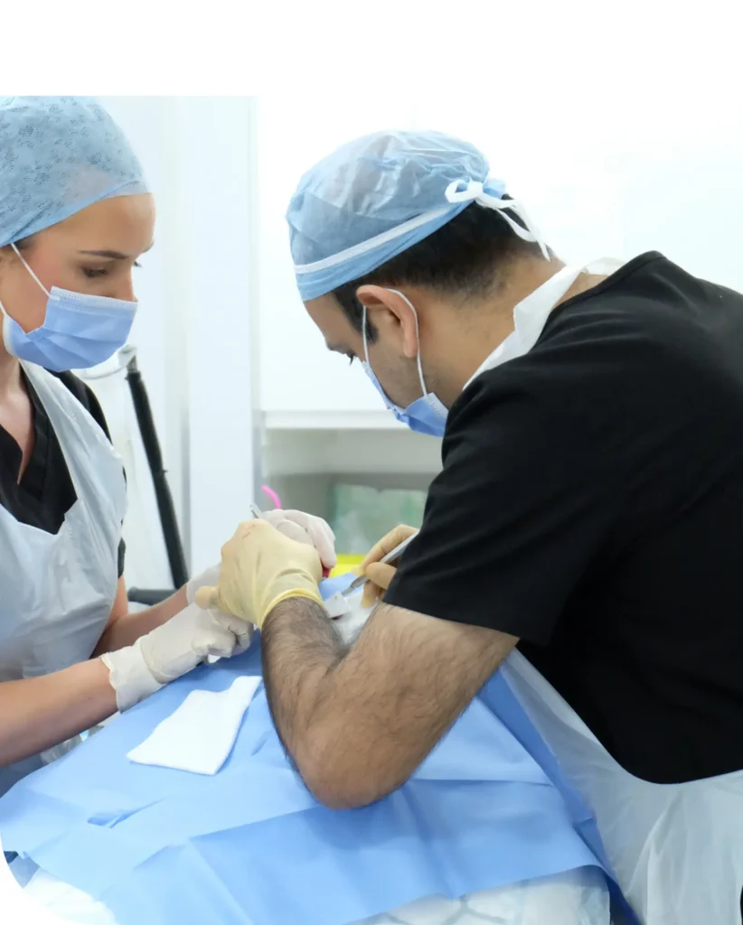
Why Is Mohs Surgery the Preferred Treatment for Nose BCC
Mohs micrographic surgery removes tumour tissue in stages, examining 100 percent of surgical margins under the microscope until no cancer remains. This method delivers:
- Cure rates up to 99 percent for primary basal cell carcinomas.
- Maximal preservation of healthy nasal cartilage and skin.
- Minimised defect size, reducing subsequent reconstruction complexity.
These benefits ensure both oncological safety and superior cosmetic outcomes on the highly visible nose.
Alternatives to Mohs Surgery
When Mohs surgery is not feasible, alternatives for basal cell on the nose removal include:
- Standard excision with 4–5 mm margins, achieving cure rates of 90–95 percent but resulting in larger defects.
- Cryosurgery for superficial basal cell carcinomas offers quick treatment but less margin certainty.
- Topical immunomodulators for very superficial lesions, sparing surgery but requiring long treatment courses.
- Radiotherapy in elderly or medically unfit patients, avoiding surgery at the expense of potential skin changes.
Each option balances convenience, cure rate and cosmetic impact, guiding individualised care decisions.
For primary lesions, the initial objective is complete tumour removal. While surgical excision and Mohs surgery are most frequently employed due to their low recurrence rates, alternative treatments may be favoured depending on the patient’s condition and tumour location.
Nasal Reconstruction After Basal Cell Carcinoma Removal from the Nose
Reconstruction restores nasal architecture and breathing function after tissue excision. Planning considers defect size, depth, and nasal subunit involvement. Effective reconstruction blends art and science to recreate symmetry, match skin texture, and preserve nasal airway integrity.
The Essential Role of Reconstruction for Function and Appearance
By closing tissue defects accurately, reconstruction:
- Preserves nasal support structures to maintain unobstructed airflow.
- Restores skin contour to minimise visible scarring.
- Protects cartilage frameworks from collapse and distortion.
Successful reconstruction prevents functional impairment and achieves natural cosmetic harmony.
Common Reconstruction Techniques After Nose BCC Removal
| Technique | Principle | Advantage | Indication |
| Primary Closure | Direct edge approximation | Fast healing, minimal scarring | Small defects (<1 cm) |
| Full-Thickness Graft | Transfer normal skin from the donor site | Good colour/texture match | Moderate defects on flat areas |
| Bilobed Flap | Two-lobed transposition flap | Preserves nasal subunit angles | Lateral tip and sidewall defects |
| Forehead Flap | Pedicled supratrochlear flap | Ample vascular supply, reliable outcome | Large tip or dorsum defects |
Each method balances vascular reliability, aesthetic match, and donor-site considerations to optimise results.
Dr Arif Aslam applies meticulous flap design, atraumatic tissue handling, and fine-suture techniques to ensure scar lines follow natural nasal contours. By aligning closures along relaxed skin tension lines and matching skin thickness precisely, he promotes inconspicuous healing and maintains nasal symmetry, delivering outstanding long-term aesthetics.

Recovery and Aftercare After Basal Cell Carcinoma Surgery on the Nose
Recovery after nose BCC removal involves staged healing, scar maturation, and ongoing protection.
A structured aftercare routine helps speed tissue repair, reduce infection risk, and support the best cosmetic results. Immediately after surgery, patients should change wound dressings daily with saline or antiseptic, take pain relief like paracetamol or ibuprofen as directed, elevate the head with cold compresses to reduce swelling, and avoid vigorous exercise or sun exposure.
Healing milestones include days 1-3 when swelling subsides, days 4-7 when dressings are removed and wounds start to close, weeks 2-4 when sutures dissolve or are removed, and months 3-6 when the scar softens and final contours appear. Following these guidelines ensures smooth recovery.
Long-term care includes applying silicone gel sheets to help collagen align properly, using broad-spectrum SPF 50+ sunscreen to prevent darkening, gently massaging the scar with moisturiser to keep it flexible, and attending follow-ups for early treatment if raised scarring develops. Consistent scar management protects cosmetic results and maintains healthy nasal skin.
Choosing Dr Arif Aslam for Basal Cell Carcinoma Removal on the Nose
Selecting Dr Arif Aslam for basal cell carcinoma removal ensures expert care backed by dual fellowship training in Mohs micrographic surgery and reconstructive dermatologic surgery, along with over a decade of experience. His precision and skill deliver outstanding oncological clearance and excellent cosmetic results.
Patients benefit from rapid access to consultation and treatment, reducing tumour growth, limiting defect size, and easing anxiety. With consistently high cure rates exceeding 99 percent and strong patient satisfaction for both function and appearance, Dr Arif Aslam offers a personalised, expert-led approach that prioritises lasting health and optimal cosmetic outcomes.
Frequently Asked Questions
Mohs micrographic surgery removes thin layers of tumour tissue one at a time, examining each under a microscope until all cancer cells are cleared. This preserves as much healthy skin as possible.
Yes, surgery creates a scar, but expert flap design and closure along natural nasal lines make scars blend in and soften with proper aftercare.
Private nose BCC removal with Mohs surgery typically ranges from £2,500 to £6,000, depending on tumour size, treatment stages, and reconstruction needs, with transparent pricing and payment plans offered.
Risks are minimal and include temporary bleeding, infection, numbness, and delayed healing, with pre- and post-operative care reducing these effectively.
Patients should provide full medical history, avoid blood thinners a week prior, arrange a companion for the day, gather post-operative supplies, and follow any fasting instructions if sedation is planned.